Magnetic bioprinting nanotechnologies for bioengineering cell-based and cell free therapies to regenerate secretory epithelia in lacrimal gland hypofunction
บทวิเคราะห์งานวิจัย
งานวิจัยนี้มุ่งพัฒนาเทคโนโลยีการพิมพ์ชีวภาพแบบแม่เหล็ก (magnetic bioprinting) เพื่อสร้างออร์แกนอยด์ (organoid) ต่อมน้ำตา (lacrimal gland, LG) เทียมสำหรับการรักษาภาวะต่อมน้ำตาทำงานผิดปกติ (lacrimal gland hypofunction) โดยใช้เซลล์ต้นกำเนิดจากเยื่อหุ้มฟัน (human dental pulp stem cells, hDPSC) เป็นพื้นฐาน งานวิจัยนี้มีความน่าสนใจและมีความสำคัญอย่างยิ่งในด้านวิศวกรรมชีวภาพและการแพทย์ฟื้นฟู เนื่องจากมีเป้าหมายในการแก้ปัญหาโรคตาแห้ง (dry eye disease, DED) ซึ่งเป็นโรคที่มีผู้ป่วยจำนวนมากและยังขาดวิธีการรักษาที่มีประสิทธิภาพ
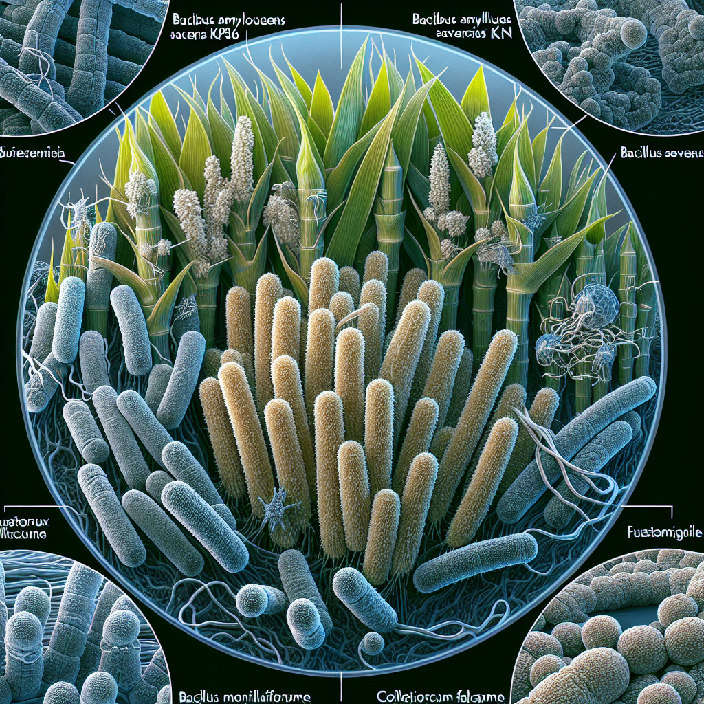
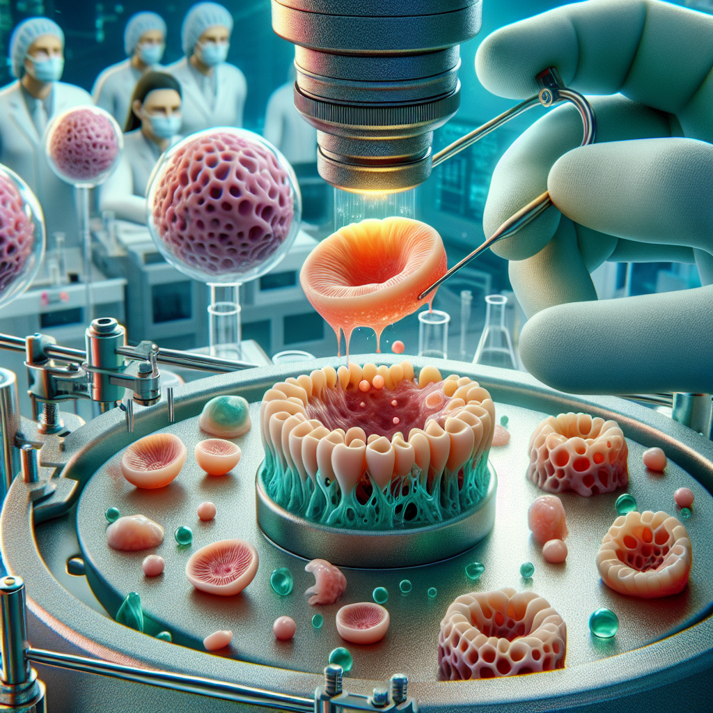
งานวิจัยแบ่งเป็นสามขั้นตอนหลัก ขั้นแรก เน้นการสร้างหน่วยการหลั่งของเยื่อบุผิวแบบสามมิติ (3D) โดยใช้เทคนิคการลอยตัวแบบแม่เหล็ก (magnetic levitation) เพื่อจัดเรียงเซลล์เยื่อบุผิวที่ได้จาก hDPSC ให้เป็นโครงสร้างคล้ายต่อมน้ำตา ขั้นที่สอง มุ่งพัฒนาการสร้างเส้นประสาท (innervation) ให้กับออร์แกนอยด์โดยการเพิ่มชั้นเซลล์ประสาท เพื่อทดสอบการตอบสนองต่อการกระตุ้นด้วยสารสื่อประสาท ขั้นตอนสุดท้าย มุ่งสร้างระบบหลอดเลือด (vascularization) ภายในออร์แกนอยด์ เพื่อให้สามารถทำงานได้อย่างยั่งยืน โดยการกระตุ้นการสร้างหลอดเลือดด้วยปัจจัยการเจริญเติบโต (growth factors) เช่น VEGF-A
จุดเด่นของงานวิจัยนี้คือการใช้เทคนิคการพิมพ์ชีวภาพแบบแม่เหล็ก ซึ่งช่วยให้สามารถควบคุมการจัดเรียงเซลล์ได้อย่างแม่นยำ สร้างโครงสร้าง 3D ที่ซับซ้อนได้ และมีความเข้ากันได้ทางชีวภาพดี นอกจากนี้ งานวิจัยยังมุ่งสร้างออร์แกนอยด์ที่สามารถทำงานได้อย่างสมบูรณ์ โดยมีทั้งส่วนประกอบของเยื่อบุผิว เส้นประสาท และหลอดเลือด ซึ่งจะทำให้สามารถศึกษาหน้าที่ของต่อมน้ำตาได้อย่างสมจริง และนำไปสู่การพัฒนาวิธีการรักษาที่มีประสิทธิภาพมากขึ้น
เป้าหมายหลักของงานวิจัยนี้คือสามประการ ประการแรก การสร้างออร์แกนอยด์ต่อมน้ำตาเทียมเพื่อใช้เป็นเครื่องมือในการศึกษาทางชีวการแพทย์ การทดสอบยา และการคัดกรองความเป็นพิษของยา ประการที่สอง การพัฒนาเทคโนโลยีการผลิตสารที่หลั่งจากเซลล์ (secretome) ในปริมาณมากเพื่อใช้ในการรักษา เช่น การใช้แบบทาภายนอกเพื่อกระตุ้นการสร้างเซลล์ใหม่ในต่อมน้ำตา และประการที่สาม การปลูกถ่ายออร์แกนอยด์เพื่อทดแทนหรือฟื้นฟูเนื้อเยื่อต่อมน้ำตาที่เสียหาย การศึกษาจะทำการประเมินคุณสมบัติทางสัณฐานวิทยา พันธุกรรม สรีรวิทยา และหน้าที่ของออร์แกนอยด์ รวมทั้งการทดสอบความสามารถในการทำงานร่วมกับเนื้อเยื่อของร่างกาย
งานวิจัยนี้มีการวางแผนการทดลองอย่างเป็นระบบ โดยแบ่งเป็นสามเป้าหมายย่อย (Specific Aims) ที่สอดคล้องกับสามขั้นตอนหลักของการสร้างออร์แกนอยด์ การศึกษาจะดำเนินการภายใต้มาตรฐานการผลิตที่ดี (current good manufacturing practices, cGMP) เพื่อให้สามารถนำไปใช้ในทางคลินิกได้ ความสำเร็จของงานวิจัยนี้จะนำไปสู่การพัฒนาวิธีการรักษาโรคตาแห้งที่มีประสิทธิภาพ ปลอดภัย และยั่งยืน และยังเป็นการก้าวสำคัญในการพัฒนาเทคโนโลยีการพิมพ์ชีวภาพแบบแม่เหล็ก ซึ่งสามารถนำไปประยุกต์ใช้กับการสร้างอวัยวะเทียมอื่นๆ ได้อีกด้วย นอกจากนี้ยังมีการพิจารณาถึงการสร้างแบบจำลองการชราภาพของต่อมน้ำตาด้วย เพื่อศึกษาผลกระทบของอายุต่อการทำงานของต่อมน้ำตา และค้นหาวิธีการชะลอหรือย้อนกลับกระบวนการชราภาพ
งานวิจัยนี้เหมาะกับอุตสาหกรรมใด
งานวิจัยนี้เหมาะกับอุตสาหกรรมหลายสาขา โดยเฉพาะอย่างยิ่ง:
-
อุตสาหกรรมยา: งานวิจัยนี้สามารถนำไปสู่การพัฒนาผลิตภัณฑ์ยาใหม่ๆ สำหรับการรักษาโรคตาแห้ง ซึ่งเป็นตลาดที่มีขนาดใหญ่และมีศักยภาพในการเติบโตสูง ออร์แกนอยด์ต่อมน้ำตาเทียมสามารถใช้เป็นเครื่องมือในการทดสอบยาใหม่ๆ เพื่อประเมินประสิทธิภาพและความปลอดภัย ก่อนที่จะนำไปทดลองในมนุษย์ การพัฒนาเทคโนโลยีการผลิต secretome ในปริมาณมากก็มีประโยชน์ต่อการผลิตยาในระดับอุตสาหกรรม
-
อุตสาหกรรมชีววิทยาศาสตร์: งานวิจัยนี้สามารถนำไปใช้ในการวิจัยพื้นฐาน เพื่อศึกษาเกี่ยวกับกลไกการทำงานของต่อมน้ำตา และการพัฒนาโรคตาแห้ง ออร์แกนอยด์ต่อมน้ำตาเทียมเป็นแบบจำลองที่มีความสมจริงสูง สามารถใช้ในการศึกษาผลกระทบของสารเคมีต่างๆ หรือปัจจัยทางสิ่งแวดล้อมต่อการทำงานของต่อมน้ำตา
-
อุตสาหกรรมเครื่องมือแพทย์: งานวิจัยนี้สามารถนำไปสู่การพัฒนาอุปกรณ์ทางการแพทย์ใหม่ๆ สำหรับการรักษาโรคตาแห้ง เช่น อุปกรณ์ปลูกถ่ายออร์แกนอยด์ หรืออุปกรณ์สำหรับการใช้ secretome ในการรักษา
งานวิจัยนี้เหมาะกับอาชีพใด
งานวิจัยนี้เหมาะกับผู้เชี่ยวชาญหลายสาขา อาทิเช่น:
-
นักวิจัยทางวิศวกรรมชีวภาพ: ผู้มีความรู้ความเชี่ยวชาญด้านการพิมพ์ชีวภาพ การเพาะเลี้ยงเซลล์ และเทคโนโลยีชีวภาพอื่นๆ เพื่อพัฒนาและปรับปรุงเทคนิคการสร้างออร์แกนอยด์
-
นักวิจัยทางการแพทย์: ผู้เชี่ยวชาญด้านโรคตา โรคตาแห้ง และการรักษา เพื่อศึกษาและประเมินประสิทธิภาพของออร์แกนอยด์ต่อมน้ำตาเทียมในการรักษา
-
นักวิทยาศาสตร์ด้านเซลล์ต้นกำเนิด: ผู้เชี่ยวชาญในการเพาะเลี้ยงและการควบคุมการเปลี่ยนแปลงของเซลล์ต้นกำเนิด hDPSC
-
เภสัชกร: เพื่อทำการคัดกรองและวิเคราะห์ประสิทธิภาพและความปลอดภัยของยาที่ใช้รักษาโรคตาแห้ง
| รหัสโครงการ : | 12554 |
| หัวหน้าโครงการ : | Joao Nuno Andrade Requicha Ferreira |
| ปีงบประมาณ : | 2563 |
| หน่วยงาน : | จุฬาลงกรณ์มหาวิทยาลัย |
| สาขาวิจัย : | กลุ่มข้อมูลด้านวิทยาศาสตร์การแพทย์และสุขภาพ |
| ประเภทโครงการ : | โครงการเดี่ยว |
| สถานะ : | ปิดโครงการ |
| คำสำคัญ : | |
| วัตถุประสงค์ : | In this project, we propose to develop in vitro bioplatforms with LG mini-organs through bioprinting organoid nanotechnologies using an MSC-line, the human dental pulp stem cells (hDPSC). As a short-term goal, we plan to engineer epithelial secretory-like units in vitro by advanced 3D biofabrication processes using magnetic levitation to spatially arrange epithelial cells (differentiated from hDPSC) as 3D acinar units. As a mid-term goal, we want to develop the innervation of the LG-like acinar units for testing response upon neurostimulation with standard neurotransmitters (acetylcholine and adrenaline analogs). These can be achieved by adding neuronal multi-layer cell sheets (e.g. Nestin+ cells) via sequential cell printing through magnetic 3D levitation. In the long-term, vascularization of the LG-like innervated acinar units is paramount to fabricate viable and more sustainable LG organoids as the final biomedical bioplatform or product. This can be accomplished by stimulating the organoids with growth factors known to induce angiogenesis (e.g. VEGF-A). In addition, this biomedical bioplaform will be further tested to evaluate the therapeutic properties of its secreted trophic factors (secretome). The ultimate goals of this project are to: (1) Create an in vitro LG-like organoid screening tool (“organ-on-a-dish”) for functional studies (towards the advancement of biomedical research) and for drug testing and cytotoxicity screening to meet pharmaceutical industry ongoing needs for drug discovery targeting therapies for LG dysfunction and DED); (2) Develop a cGMP-based technology to produce hDPSC-derived secretome in large scale (large quantities) for in vivo use. For example, the topical delivery of secretome (cell-free therapies) could potentially stimulate LG regeneration in in vivo epithelial injury models; (3) In vivo transplantation of epithelial organoids (cell-based therapies) for partial or total replacement or regeneration of epithelial secretory tissue damaged in LG hypofunction models. 11.1. Hypothesis and Specific Aims To address the critical need to fabricate in vitro LG organoids for both the biomedical research/industry and in vivo/clinical use, we hypothesize that LG organoids developed by 3D levitation nanotechnology can generate functional and sustainable LG-like secretory organoids with innervation and vascularization, comparable to the native LG organ. To test this central hypothesis, we propose three Specific Aims (SA): SA #1: Identify the morphological, genotypic, phenotypic and functional properties of acinar epithelial units from LG organoids developed by 3D magnetic levitation. This can be accomplished by using biocompatible nanoparticles to magnetize hDPSC-derived epithelial cells to rapidly create 3D cell rings mimicking native architectures of LG acinar epithelial units. Core acinar epithelial units will be formed by 3D cell printing and magnetic levitation techniques. Moreover, these 3D structures can be subjected to 3D automated image acquisition to evaluate for growth, size, shrinkage and function in vitro. All these protocols can be performed under cGMP (current good manufacturing practices) conditions for clinical applicability. Morphological, genotypic, phenotypic and functional assays will be performed to fully characterize these organoids in vitro. Organoid viability (at cellular level) and cytotoxicity testing will also be performed after transplanting these epithelial organoids into an ex vivo organ model. SA #2: Determine the presence and viability of neuronal populations and the response to neurostimulation of LG-like organoids. This will be accomplished after the assembly of neuronal cell sheets into acinar epithelial core units of the LG organoids. SA #3: Characterize the vascular network and the integration of the final LG organoid after transplantation into a LG injury model. This step is essential to understand the vascularization and cell viability and sustainability of the organoid after transplantation. Integration and communication of the different tissue compartments of the LG organoids (epithelial, neuronal, vascular) with the host LG will be assessed. Regenerative paracrine signals produced by the LG organoid will also be evaluated. Epithelial fetal LG organ models from mice will be used to test the epithelial regenerative ability in ex vivo conditions. |
Joao Nuno Andrade Requicha Ferreira. (2563). Magnetic bioprinting nanotechnologies for bioengineering cell-based and cell free therapies to regenerate secretory epithelia in lacrimal gland hypofunction. จุฬาลงกรณ์มหาวิทยาลัย. .
Joao Nuno Andrade Requicha Ferreira. 2563. "Magnetic bioprinting nanotechnologies for bioengineering cell-based and cell free therapies to regenerate secretory epithelia in lacrimal gland hypofunction". จุฬาลงกรณ์มหาวิทยาลัย. .
Joao Nuno Andrade Requicha Ferreira. "Magnetic bioprinting nanotechnologies for bioengineering cell-based and cell free therapies to regenerate secretory epithelia in lacrimal gland hypofunction". จุฬาลงกรณ์มหาวิทยาลัย, 2563. .
Joao Nuno Andrade Requicha Ferreira. Magnetic bioprinting nanotechnologies for bioengineering cell-based and cell free therapies to regenerate secretory epithelia in lacrimal gland hypofunction. จุฬาลงกรณ์มหาวิทยาลัย; 2563. .
Creative Commons : CC

รายการที่เกี่ยวข้อง
OECD : กลุ่มข้อมูลด้านวิทยาศาสตร์การแพทย์และสุขภาพ